Single biological systems
- Contact : Karen Perronet
Objectives
Our goal is to characterize the real-time dynamics of biological processes at the single-object level. By measuring many individual systems, we can determine not only average parameters (such as speeds, interaction times, etc.) but also the statistical distribution of these parameters—without needing to synchronize the systems. Moreover, if a subpopulation behaves differently, we can detect it. Its behavior won’t be "drowned out" in the overall measurement!!
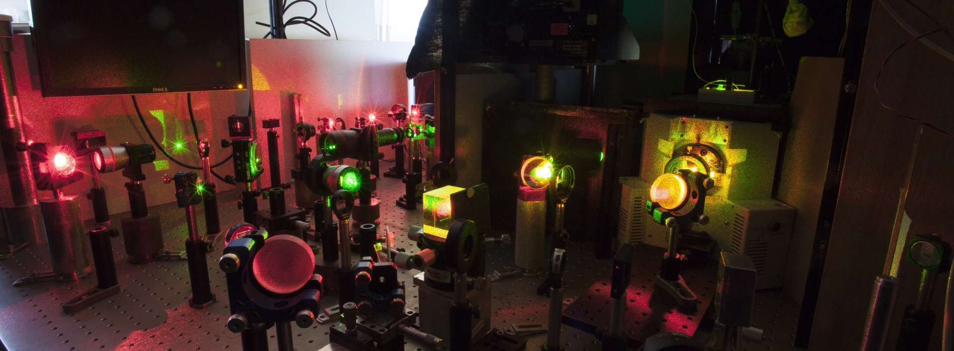
Methods
To achieve this, we use advanced microscopy techniques. The first is Total Internal Reflection Fluorescence Microscopy (TIRFM), a dark-field fluorescence method that isolates signals from individual fluorescent molecules. We use this technique to specifically label the biological samples we study.
We also develop Interferometric Scattering Microscopy (iSCAT), which records the interference signal between a reference beam and the beam scattered by a molecule. This allows us to determine the molecule’s mass.
Main collaborators
Olivier Namy (I2BC, Gif sur Yvette)
Guillaume Tresset (LPS, Orsay)